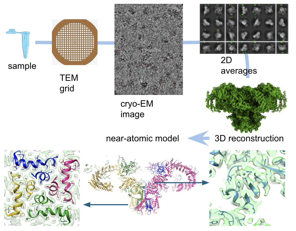
The 1,300 square foot cryo-electron microscopy (cryo-EM) facility is in a newly renovated space in the basement of VCU’s McGlothlin Medical Education Center, at 1201 E. Marshall St, Richmond, VA. The facility provides investigators with the capability to carry out cryo-EM and 3D reconstruction of proteins and macromolecular complexes up to high resolution.
The facility is endowed with a Thermofisher Scientific Tundra cryo-electron microscope and all necessary ancillary equipment: a Vitrobot Mark IV cryo-plunger, a Pelco easyGlow glow discharge unit, a Denton DV 502B evaporator, a bench for negative staining, and a bench for cryo-grid loading. The facility also has a linux server with 250 TB storage and four 20 GB GPUs with image processing software (cryosparc, spider, phenix, coot, chimera, chimerax, etc). The equipment was purchased using Virginia General Assembly Higher Education Equipment Trust Fund and VCU funds.
The goal of cryo-EM is to obtain the three-dimensional structure of purified macromolecular complexes such as proteins, protein-DNA, protein-RNA, and protein embedded in small liposomes. In addition to 3D density maps at different levels of detail up to near-atomic resolution, cryo-EM data reveals other important structural information such as location of ligands, subunit organization, conformational landscape, and identification of regions of variability.
The cryo-EM facility operates as a full-service core, performing sample preparation for negative staining and cryo-EM, high throughput data collection, and image processing. The facility will also provide training on all these operations to enhance access and user capability.
In addition to providing service to the VCU community, we also offer cryo-EM services to outside organizations. Please contact us for your cryo-EM needs and/or potential collaborations.
To cite the facility in your manuscript, use its unique Core Facility Research Resource Identifier: RRID:SCR_027178
Services
- Discussion on feasibility
- Negative staining sample preparation
- Cryo-EM sample preparation
- High throughput cryo-EM and negative staining data collection
- On-the-fly preliminary 3D structural determination (simultaneous to data collection)
- Image processing (2D averaging, 3D reconstruction, 2D and 3D classification, etc.)
- 3D structure rendering and interpretation, model building
- Training on negative staining, cryo-EM sample preparation, data collection, image processing, model building, and rendering of 3D structures.
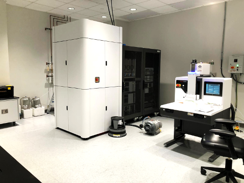
Tundra cryo-Em (left); Sample loading station (right)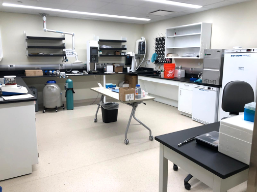
Sample preparation room
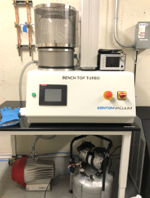
Evaporator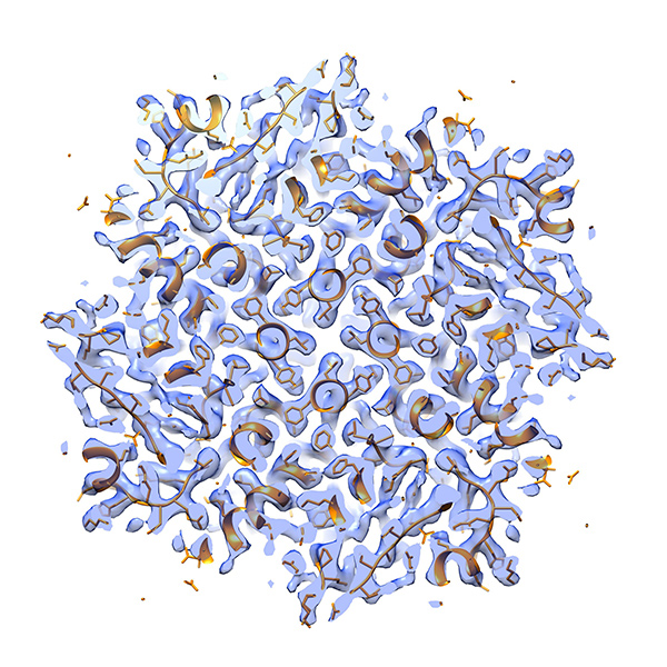
Apoferritin at 3 Å resolution
(obtained at the cryo-EM Facility)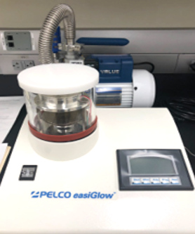
Glow discharge apparatus
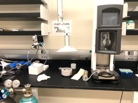
Virobot Mark IV cryo-plunger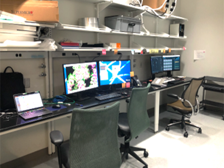
Computer room
Location
Cryo-Electron Microscopy Facility
MMEC Building
Suite B-106, rooms B-E
1201 E. Marshall St.
Richmond, VA 23298
Contact us
Montserrat Samso, Ph.D.
Co-director, Structural Biology core
Department of Physiology and Biophysics, School of Medicine

Alimohammad Hojjatian, Ph.D.
Manager, Cryo-Electron Microscopy facility, Structural Biology core

