Descriptions
Molecular imaging is a multidisciplinary science that provides non-invasive visual characterization and measurement of the internal cellular functions and molecular processes of humans and other living organisms in their intact physiological environment.
Magnetic resonance imaging and spectroscopy
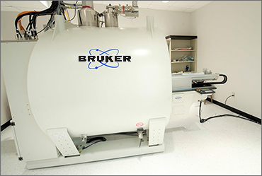
The 7T/30 preclinical MRI system is a 7 Tesla 30 cm bore MR scanner (manufactured by Bruker Biospin). It is a multipurpose research system for high-resolution magnetic resonance spectroscopy and imaging in selected volumes and 2- or 3-dimensional image reconstruction based on the principles of nuclear magnetic resonance.
- 7.0 Tesla (300 MHz) Bruker Biospec system
- 2 independent gradient inserts for flexibility among sample size; 200 and 120 mm inner-diameter, capable of gradients strengths of 300 mT/m and 600 mT/m, respectively
- 2 independent RF transmitter channels, capable of 1000-watt output each
- RF coils for proton imaging and spectroscopy with inner diameters of 154, 86 and 40 mm
- Actively decoupled, 4-channel receive only coils for rat and mouse cranial imaging
- Dual tuned volume coil for proton/fluorine imaging and spectroscopy
- Dual tuned surface coils for proton/phosphorus and proton/carbon imaging and spectroscopy
MultiScan LFER large bore research PET/CT
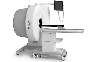
MultiScan LFER large bore research PET/CT is a multi-use preclinical PET/CT imaging system. The unique and patented design of the MultiScan LFER (Large Field of view Extreme Resolution) features a large bore with 20 cm axial and 15 cm transaxial field of view (FOV). This feature, together with quad mouse chamber, allows imaging of multiple mice or imaging of other experimental animals larger than mice and rats. The MultiScan LFER PET/CT with sub-mm PET and CT resolution with integrated animal support system is useful for a wide range of translational imaging applications. This system features the most versatile preclinical PET/CT gantry ever built. The system can deliver PET images with very high resolution and high sensitivity (5%) at the same time. The spatial resolution can reach 1 mm with proprietary PET 3D iterative reconstruction methods. Efforts are also underway to incorporate a quad mouse chamber with the LFER PET/CT that would make high-throughput preclinical PET/CT research imaging a possibility resulting in significant reduction of cost for each experiment that uses vendor supplied PET tracers.
Optical imaging
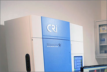
CRi Maestro-2 is a multispectral small animal imaging system (originally manufactured by CRi Inc, USA - presently owned by Perkin Elmer, USA). The Maestro-2 contains an optimized, solid-state liquid crystal tunable filter (LCTF) based multispectral imaging camera that can capture high-spectral-resolution multispectral fluorescence images of small animals at multiple, user-defined wavelengths ranging from the blue out to the near infrared. The hardware is complemented by state-of-the-art algorithms for accurate spectral unmixing that allow the user to subtract the background autofluorescence, thereby improving the target/background ratio to generate high-contrast optical images from fluorescent signals.
The CRi Maestro 2 is a high-performance multispectral imaging system designed for fluorescence macro-imaging of small animals such as mice or rats.
The system is equipped with unique multispectral tunable optics and inbuilt multispectral imaging software algorithms which enables users to rapidly image and quantify more than one fluorescent signal within a mouse or rat in real time.
The system can be used for multiplexing and spectral unmixing enabling imaging of multiple fluorescent signals simultaneously together with the removal of any autofluorescence from an image. Spectral Unmixing and Dynamic Contrast Enhancement (DyCE™) allows quantification of temporal biodistribution of fluorescent markers at much earlier time points (first few seconds following injection).
Other features:
- In vivo fluorescence imaging
- In vivo multispectral imaging and unmixing
- Autofluorescence correction
- Minimum image pixel resolution 25 micron
- Heating stage and anesthesia manifold
- Solid-state liquid crystal tunable filter
- Imaging wavelength range from 500 to 950 nm
- 3 mice could be imaged simultaneously
Photoacoustic imaging
The iTheraMedical MSOT inVision optoacoustic imaging platform for small animal imaging. The unit can acquire cross-sectional images, with high resolution, in vivo and in real time. The unique capability of the MSOT inVision to visualize and quantify optoacoustic contrast throughout the whole animal, from head to tail, makes it a very attractive choice for many research applications.
The MSOT inVision features 360˚ ring illumination and 270˚ acoustic detection. This wide coverage angle is the basis for the full cross-sectional views of small animals and the unmatched tomographic image quality.
Multispectral optoacoustic tomography (MSOT) system (iThera Medical GmBH, Munich, Germany). The MSOT is a novel technology providing tomographic optoacoustic/photoacoustic in vivo identification of spectral signatures from multiple specific agents, along with excellent intrinsic tissue contrast. This system is based on the photoacoustic effect. In 1881, Alexander Graham Bell discovered that light energy absorbed by a material results in an acoustic signal. Modern optoacoustic imaging systems such as the MSOT use high-energy pulsed lasers and highly sensitive broadband ultrasound detectors. By exciting tissue with a laser pulse and detecting the signals generated by the conversion of light energy into sound waves, optical absorption in tissue can be detected and visualized. Endogenous chromophores in the NIR include oxy-hemoglobin, deoxy-hemoglobin, melanin, lipid and collagen. Extrinsic absorbers include fluorophores, quenchers, various kinds of nanoparticles and other probes used for optical imaging.