BARC core
Our mission is to serve as a central resource to help scientists discover how utilizing research registries at VCU can advance their scientific achievements, in conjunction with providing excellent support services to see projects through from nascent stage to data analysis.
About
The Bioimaging and Applied Research Core, directed by Frank Corwin, Ph.D. and managed by Mackenzie Newman, Ph.D., offers comprehensive instrumentation and technical support for all aspects of in vitro and in vivo pre-clinical non-human anatomical and molecular imaging. Studies may be conducted on samples prepared to exhibit characteristic features for a particular imaging modality. Within the intact environment, physiological and pathophysiological activity may be observed through imaging or numerous biological pathways and interactions involved in injury and disease progression, such as traumatic brain injury, cardiac disease, tumorigenesis, and therapeutic interventions.
BARC offers a wide variety of services via a traditional fee-for-service model and collaborative solutions that involve do-it-yourself imaging and data analysis and lab-expertise for hire. These models give investigators the opportunity for training and hands-on time on selected instrumentation and permit investigators to purchase technician time from experienced staff to contribute to protocol development and optimization of new approaches. Staff can assist in experimental planning and project design.
News
4/23/26: Congratulations to the Spiegel group for their recent Nature Communications publication, "SPNS2 exports sphingosine-1-phosphate and imports glucose" (PubMed link; doi: 10.1038/s41467-026-71659-7, including data generated in BARC! We are proud to see this novel work out and look forward to what future studies it influences.
4/15/26: Thanks to everyone who joined us at the Preclinical Imaging Consortium 2026 meeting in Philadelphia! It was great to exchange ideas with our fellow cores to improve our research. We look forward to next year!
4/1/26: Congratulations to the Garcia-Bonilla/Limbrick group for their new publication, "Post-hemorrhagic hydrocephalus of prematurity is associated with disruption of tight junctions and increased macrophage activity in the choroid plexus" (PubMed link; doi: 10.1186/s12987-026-00800-x). BARC is thrilled to have been a part of the study and are excited to see where future studies go!
ANNOUNCEMENT: BARC is now offering fabrication and object duplication services! Using our advanced imaging equipment and high-resolution resin 3D printers, we are able to produce functional duplicates of most small objects at <100μm resolution. Models can be printed directly from 3D scans, traced from 3D scans to correct imperfections, or generated from the ground up. Alternate fabrication methods such as vacuum/thermoforming, silicone casting, laser cutting, and more are available. Rates start around $50 and can be charged to any index code. Please contact us for more information!
Initiate a project
BARC offers a new business model based on a broad range of off-the-shelf, state-of-the-art services as well as the ability to develop and tailor protocols for specific research projects. To initiate a service request or to schedule a discussion about an ongoing or upcoming project please use our intake form or contact us via email.
Acknowledge the VCU BARC Core
Published work using data generated at BARC should include the following in the acknowledgment:
The data included in this study was generated at the Bioimaging and Applied Research Core facility at Virginia Commonwealth University.
Suggested grant language
Include the following text in the Facilities and Resources section of grant applications when proposing to conduct work that utilizes the BARC facility:
The Bioimaging and Applied Research Core laboratory (BARC) is a VCU institutionally-designated shared resource providing state-of-the-art imaging technologies to support research in life and physical sciences, with an emphasis on multi-modality imaging approaches to study structure, biology, biochemistry and pharmacology in vitro and in vivo.
The BARC is directed by Frank Corwin, Ph.D., and managed by Mackenzie Newman, Ph.D., who have more than 30 years of combined experience in the field of non-invasive in vivo imaging. The BARC provides a hub for translational research, with molecular imaging equipment that supports imaging of small mammals, as well as supporting physical sciences applications such as fluid dynamics.
Our shared resource facility for anatomical and molecular imaging increases the ability of investigators to translate preclinical concepts of injury and/or cancer into detection, therapy, and remission in animal model systems and, ultimately, into early phase clinical trials. Further, it provides expertise and instruments that facilitate direct, specific, and ultra-sensitive studies of molecular pathways in the in vivo environment. This always-expanding core leverages multiple imaging technologies, combined with vendor-supplied imaging probes and contrast agents, available through VCU BARC.
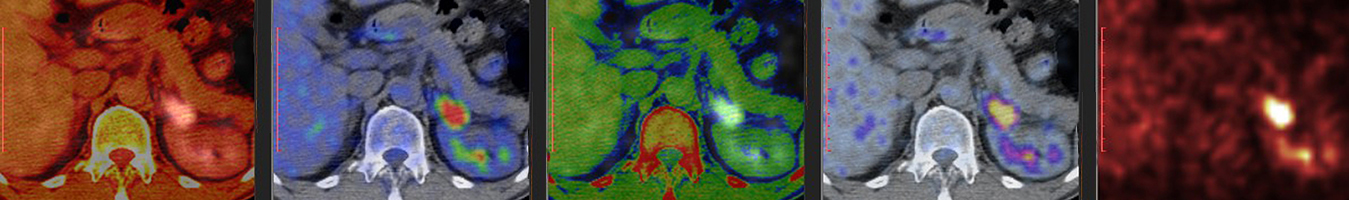
The BARC offers radionuclide imaging services of small animals on PET as well as non-radionuclide-based imaging using MRI/MRS, photoacoustic, and multispectral fluorescence systems. The facility also provides project consultation, user training, and staff-assisted data analysis services.
Located on the VCU MCV campus, the BARC maintains a significant portfolio of instrumentation in support of in vitro and in vivo imaging. The state-of-the-art imaging equipment includes:
- Bruker Biospec 7 Tesla preclinical magnetic resonance imager system, with multinuclear capability
- LFER PET/CT in-vivo animal imager (Mediso USA), capable of large field-of-view (15 cm axial, 20 cm trans-axial), whole body preclinical imaging
- Multispectral optoacoustic tomography (MSOT) system (iThera Medical GmBH, Munich, Germany)
- Maestro-2 multispectral small animal imaging system (CRi Inc).
Commonly provided services include:
- Training on the operation of the imaging modalities
- Direct operations and data analysis support on the more complex modalities
- Project design and implementation guidance
Additionally, BARC has immediate access to separate animal holding facilities available adjacent to the imaging suites, which are approved to house animals injected with radioactive substances. Sufficient space for housing mice and rats is available and includes cages with filter tops, laminar flow racks, and biosafety cabinets to prevent the spread of infectious disease. The VCU Division of Animal Resources provides animal care 365 days a year. Veterinary consultation services are readily available, as well as training and technical services regarding surgery and blood sampling.
Recent citations
Weigel, C., Hossen, M.L., Brown, R.D.R. et al. SPNS2 exports sphingosine-1-phosphate and imports glucose. Nat Commun (2026). https://doi.org/10.1038/s41467-026-71659-7
Garcia-Bonilla, M., Swarup, R., Limbrick, O. W., Vohra, H. Z., Otun, A., McKalip, K., Bernhardt, W., Shumilov, K., Michenkova, M., Crouthamel, J., Newman, M., Dikranian, K., McAllister II, J. P., & Limbrick Jr, D. D. (2026). Post-hemorrhagic hydrocephalus of prematurity is associated with disruption of tight junctions and increased macrophage activity in the choroid plexus. Fluids and Barriers of the CNS. https://doi.org/10.1186/s12987-026-00800-x
Hawkins IK, Phi T, Mitchell J, et al. <scp>MYH6</scp>‐Cre Insertion Accelerates Cardiac Phenotype in Dystrophic D2‐mdx Mice. The FASEB Journal. 2025;39(14). doi:10.1096/fj.202500792r
C. Weigel, Md L. Hossen, R.D.R. Brown, C. E. Senkal, C.D. Green, J. Newton, S. Saha, E.N.D. Palladino, B. Ni, F.S. Celi, X. Fang, F.D. Corwin, H.Z. Li, D.B. Sauer, P.P. Chapagain, and S. Spiegel. “SUGAR FOR THE SPHINX: SPINSTER HOMOLOG 2 TRANSPORTS SPHINGOSINE-1-PHOSPHATE OUT AND GLUCOSE IN CELLS”. FEBS special meeting “Sphingolipid biology: breaking boundaries”. 25-30 May 2025.
Brøndsted, F., McAfee, J.L., Moore, J.D. et al. Acoustic loudness factor as an experimental parameter for benchmarking small molecule photoacoustic probes. Nat Commun 16, 3779 (2025). https://doi.org/10.1038/s41467-025-59121-6
Wiggins, C.S., Cabral, A., Mafi, A. et al. Integrated positron emission particle tracking (PEPT) and X-ray computed tomography (CT) imaging of flow phenomena in twisted tape swirl flow. Exp Fluids 65, 121 (2024). https://doi.org/10.1007/s00348-024-03860-7
India K Hawkins, Josh Mitchell, Adolfo G Mauro, Nigeste M Carter, Frank Corwin, Fadi N Salloum, Frank J Raucci. "Measuring Fibrosis Progression in Duchenne Cardiomyopathy Using Cardiac Magnetic Resonance in mice". Basic Cardiovascular Sciences Conference, 22-25 July 2024.
Location
1101 E Marshall St
Sanger Hall
Rm. B3-027
Richmond, VA 23298
Contact us
Questions about core laboratories at VCU? Please email: